Review Article

Biochemical and Molecular Studies of Various Enzymes Activity in Fungi  

Muhammad Ishfaq1 , Nasir Mahmood2 , Qurban Ali3 , Idrees Ahmad Nasir3 , Muhammad Saleem1
1 Molecular Genetics Research Laboratory, Department of Botany, University of the Punjab (New campus), Lahore, Pakistan
2 Department of Human Genetics and Molecular Biology, University of Health Sciences, Lahore, Pakistan
3 Centre of Excellence in Molecular Biology, University of the Punjab (New Campus), Lahore, Pakistan
Author    Correspondence author
Molecular Plant Breeding, 2016, Vol. 7, No. 9   doi: 10.5376/mpb.2016.07.0009
Received: 07 Dec., 2015    Accepted: 15 Jan., 2016    Published: 15 Jan., 2016
© 2016 BioPublisher Publishing Platform
This is an open access article published under the terms of the Creative Commons Attribution License, which permits unrestricted use, distribution, and reproduction in any medium, provided the original work is properly cited.
Preferred citation for this article:

Ali Q., Ishfag M., Mahmood N., Nasir I.A., and Saleem M.. 2016, Biochemical and Molecular Studies of Various Enzymes Activity in Fungi, Molecular Breeding, 7(9): 1-16 (doi: 10.5376/mpb.2016.07.0009)

Abstract

Fungi are decomposers in most ecosystems and make important contribution to the ecological balance of our world. They have great industrial importance due to the presence of different enzymes like laccase, superoxide dismutase, cellulases, amylases and catalase etc. These enzymes performed synthetic and degradative functions. They are physiologically necessary for all of the living organisms and are universally occur with wide genetic diversity in plants, animals and micro-organisms. Mostly the micro-organisms are an attractive and efficient source of various enzymes and also owing to have the limited space required for their cultivation and their ready susceptibility to genetic manipulations. Although the extensive research on various aspects of these enzymes, there is scarcity of the knowledge about the role that governed the diverse specificity of these enzymes. After deciphering the secrets about these enzymes would enable us to exploit their use in biotechnology. Fungi have vital roles in biotechnology such as production of drugs and enzymes. Fungi can be cultured easily and hence they can be used in microbiological, genetic and molecular research. It is very important to investigate genes and the role of genes that are responsible for the formation of these enzymes. In the current review described the study of production of laccase, superoxide dismutase and catalase enzyme through various fungi, the activity of enzymes and the genetic diversity of genes involved for the formation of these enzymes.

Keywords
Laccase; Superoxide Dismutase; Cellulases; Amylases; Catalase; Enzymes; Fungi

1Introduction
Fungi have been used in modern scientific research due to their high potential for different enzymes production based on genomic features. The great proportion of soil mycoflora represented by saprobic fungi plays an important roles in decomposition, thus contribute to the global carbon cycle (Nevalainentics and Penttila, 2003). Different catabolic activities by different enzymes enable fungi to colonize on organic matter and this feature explains the significant use of filamentous fungi in biotechnology (Kaeberlein et al., 2002). Filamentous fungi secrete different enzymes in the growth medium, and most of these enzymes are hydrolytic in nature and employed in different industrial processes (Shakuntala et al., 2009). There is need to characterize fungal strains at molecular level for efficient enzyme production (Gerd et al., 2006). Sordaria fimicola (Roberge ex Desm.) Ces. & De Not., is an important fungus belonging to Ascomycota. Sordaria fimicola is a filamentous fungus growing as extending and branching tubular cells (hyphae) that generally grow radially with symmetric colony (Alexopolous et al., 1996; Kavak, 2012). The natural habitats of S. fimicola have been mainly defined in dung of herbivorous animals (Masunga et al., 2006). However, it has been isolated from different habitats including maize stalks (Alma et al., 2000). Colonies of S. fimicola grow rapidly on Potato Dextrose Agar (PDA) medium in 7 days at 28ºC reaching 9 cm in diameter, from brown to dark brown mycelium with homothallic perithecia (Jeamjitt, 2007).
 
The S. fimicola strains were first time isolated from “Evolution Canyon I (EC I)” (Nevo, 1995) located at mountain Carmel, Israel were included (Figure 1). The south facing slope (SFS) of EC I has dry and harsh environment with quite different flora and fauna as compare to north facing slope (NFS) which has moist and lush green environment. The age of EC 1 is almost 3 to 5 million years (Nevo, 1997). The north and south facing inclines are 100 and 400 meters apart from each other from bottom and top respectively (Pavlicek et al., 2003). It was proposed that mutation and genetic diversity was more frequent in S. fimicola strains belonging to SFS as compared to NFS of EC I (Nevo, 1995, 1998). Genomic DNA is affected by environmental changes which are the basis of biodiversity studies; therefore, the scientific aspect of biodiversity needs to be explored (Saleem et al., 2001). On the both slopes pedology, geology (Karcz, 1959; Nevo et al., 1998) and regional climate (mediterranean) is same with annual rainfall approximately 600 mm, 1700 mm evapo-transpiration rate and mean temperature for January and August is 13 and 28°C, respectively (Atlas of Israel, 1970). Due to differences in geographic orientations, south facing slope receives more radiations annually as compare to northern slope (Kirkby, 1990) and has savannoid vegetations, while northern slope has dense evergreen and deciduous trees (Nevo et al., 1999). According to Pavlicek et al., (2003) the African south facing slope receives 200-800 % more solar radiations than the European north facing slope. One S. fimicola strain (SF13) included in current research was obtained from Miller’s Mycological Lab. UIUC, isolated from the surroundings of University of Illinois at Urbana-Champaign, USA. The Urbana-Champaign city has humid continental climate having four distinct seasons with hot summers (32.2°C) and cold winters (-17.8°C).


 Figure 1 Picture of ‘Evolution Canyon I’ Mount Carmel, Israel with assigned stations. The green lush European’ temperate, cool-mesic north facing slope (NFS) sharply contrast with warm-xeric, tropical, ‘African’ south facing slope (SFS). Adopted from (Raz et al., 2009)


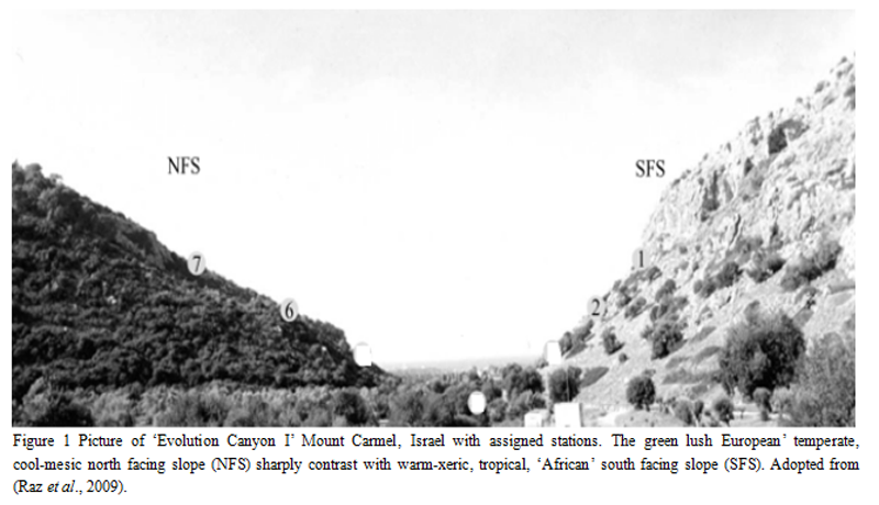
Lamb et al., (2008) worked on the crossing over, gene conversion and variation in recombination properties of S. fimicola wild strains isolated from opposite slope of EC 1 and reported that recombination frequencies were higher in south facing slope than that of north facing slope where conditions are mild. The hypothesis that mutation rate would be less in natural strains taken from north facing slope, which has moist and lush green environment than those from the harsh and stressful environment of south facing slope was proposed by Nevo, (1995) which needs to be further explored. Variations in genome induced as a result of stress have been found in several other organisms including Drosophila melanogaster, A. mystacinus (Nevo et al., 1998), S. fimicola (Rottenberg et al., 2006) and N. linckia (Dvornyk and Nevo, 2003). According to Nevo, (1997) 9 out of 14 model organisms exhibited higher genetic diversity which belongs to more harsh and heterogenous SFS. Mutation frequencies, DNA repair, gene conversion, genetic recombination, SNP, retrotransposons and genetic diversity was found higher at more stressful SFS (Nevo, 2001). Three fold higher rates of heritable mutation in S. fimicola, a coprophilous fungus and 4 fold higher of genetic recombination have been found in D. melanogaster on stressful heterogenous SFS as compared to mild- moist NFS (Nevo, 1997).

 
1 Genomic Diversity Analysis by RAPD Analysis
Study and investigation of variations in genes can be helpful in understanding various phenomena at molecular level (Hibbett, 1992). Random amplified polymorphic DNA analysis can be helpful in identifying genomic modifications among organisms of even same species due to high variability of markers (Sunnucks, 2000; Nawaz et al., 2013). Increased numbers of genetic characters lead towards studying the diversity of fungal species (Ellstrand and Roose, 1987). There are two types of basic markers, PCR and non-PCR based, which are easy to use with reliability, precision of analysis, statistical influence and confidence of revealing polymorphism in genes and genomes (Agerwal et al., 2008). Molecular markers which are PCR based are excellent tools for defining relationships in fungi at genetic level (Welsh and McClelland, 1990; Duran et al., 2009). The significance of PCR-based marker methods is due to that it is rapid and needs small amount of genomic DNA (Jacobson and Hedren, 2007). Random amplified polymorphic DNA analysis first applied on genomic diversity analysis was based on the use of oligonucleotide primers and genomic DNA (Williams et al., 1990). Genomic variations of different fungal species can be studied by using RAPD markers (Crowhurst et al., 1991). Random amplified polymorphic DNA technique involved the availability of priming site on whole genome for a single primer in inverted position and closes enough to allow PCR amplification (Whitekus et al., 1994). The short primers optimized for RAPD analysis have become genomic markers which can be used for quantitative assessment of genomic similarities of strains from same or different species of organisms (Leung et al., 1992).
 
In addition to RAPD several other marker systems are being applied for exploration of biodiversity including RFLP, SSR and AFLP (William and Clair, 1993; karp et al., 1997). According to Nesbitt et al., (1995) RAPD markers can be used in paternity analysis, taxonomic-based identification and genetic diversity (Van de Ven and McNicol, 1995). Random Amplified Polymorphic DNA analysis is being used for generating genomic maps (Tulsieram et al., 1992) and in detecting loci of interest for studying molecular biodiversity in fungi (Plomion et al., 1996). Random Amplified Polymorphic DNA markers are cheap and utilize little amount of genomic DNA for analysis (Soares et al., 2008). Random Amplified Polymorphic DNA markers analysis is more reliable in terms of reproducibility and provides high resolution of genotype distribution in natural populations (Brahmane et al., 2008). Therefore, RAPD has been a very popular molecular technique to generate genus-specific, species specific or strain-specific diagnostic DNA fragments or fingerprints, identifying genes linked to traits of interest; undertaking genetic diversity studies and gene mapping for development of diagnostic and identification of living organisms (Bazzicalupo and Fani, 1996; Abad et al., 1998; Ransom et al., 1998). Random Amplified Polymorphic DNA is also being used in population genetics studies like genetic diversity, divergence within and among populations based on assumption of Hardy-weinberg equilibrium (Brown and Epifanio, 2003). It detects the genetic variations in the genome of an organism in terms of sequence variation at the priming regions (Magalhães et al., 2007). Similarities in banding profiles among strains (i.e. the number and sizes, but not the intensity of amplified bands) can be calculated and used to infer strain relationships (Dutra et al., 2008). Random Amplified Polymorphic DNA technique can also be applied to estimate the populations, for which no particular molecular markers have been established, thus facilitating the screening process of genetic variability (Lacerda and Wrobel, 2001). Due to nucleotide sequence differences either by insertions or deletions in the fragment size between two primer sites, length differences occur that may lead to polymorphism which is related to genomic diversity (Agerwal et al., 2008).
 
Shah et al., (2006) differentiated the A. niger genome by using RAPD and the 0.7 kb fragment amplified was used to further differentiate from other strains of A. niger (ATCC 16880). Genetic relationship among A. niger, Aspergillua flavus and Aspergillus parasiticus were determined by RAPD analysis (Swelim, 2005a) which revealed 37, 59, and 51 % polymorphism, respectively. Furthermore, RAPD analysis demonstrated that genetic similarity was 37 % in A. niger, 58 % in A. flavus, and 51.5% in A. parasiticus (Aiat, 2006). Random Amplified Polymorphic DNA analysis was used in detecting genetic diversity in many phytopathogenic fungi (WÖstemeyer and Kraibich, 2002; Sharma et al., 2002; Sharma, 2003). Due to high variability RAPD markers can be used to detect differences within and among species at genomic level (William et al., 1990; Parker et al., 1998; Sunnucks, 2000) as well it is also helpful to explore intra-specific variations in large number of fungi on genetic bases (Fegan et al., 1993; Moore et al., 2001).
 
1.1 Ribotyping
The 18S rRNA gene has been used to characterize fungal strains at species level (Meyer et al., 2010). Phylogenetic analyses of fungal taxa at different levels can be done by using 18S rRNA gene that is considered as phylogenetic marker. In all living organisms ribosomal ribonucleic acid (rRNA) is involved in protein synthesis and it comprises of 90 % of total RNA (Forster and Toth, 2003). The ribosome has two subunits smaller subunit (SSU) and large subunit (LSU) (Higgs, 2000). The LSU acts as ribozyme, which catalyzes the peptide bond formation. All SSUs have one large ribosomal subunit molecule termed as 16S in Archaea, Bacteria and 18S in Eukaryotes (Moore, 2009). The 18S rRNA gene consists of ~1900 nucleotides and responsible for the translation of different proteins. The sequences of 18S rRNA genes are widely used to find out evolutionary relationship among different organisms (Smit et al., 2007). The flanking region of the 18S rRNA gene is highly conserved and has been used as a reliable marker to determine environmental biodiversity in the species of different organisms (Woese et al., 1990; Hanif et al., 2012). To segregate fungi into diverse strains within species amplification of rRNA gene for ribotyping and SNPs analysis has become essential molecular aspect (Balajee et al., 2008). Molecular systematics is an important tool in recent taxonomy of fungi (Bruns et al., 1991, Mitchell et al., 1995). DNA sequence data of 18S, 26S, ITS (Internal Transcribed Spacer) along with mitochondrial rDNA are abundantly used in current phylogenetic studies in case of eukaryotic cells (Wilmotte et al., 1993; Shan et al., 2015; Zameer et al., 2015). Due to conserved nature of 18S rDNAs they are applied in phylogenetic analyses of higher taxonomic rank fungi (Swann and Taylor, 1993). The advent of molecular phylogenetic and the use of ribosomal RNA (rRNA) as a molecular chronometer extended phylogenetic studies to different organisms including the microbial world where it was difficult to find distinguishable, observable phenotypes and resulted in the classification of life into a tripartite world (Woese et al., 1990). A number of insertions and deletions have been reported in variable domains of rRNA gene including V2, V4, V6, 8, and V9 domains (Bruns et al., 1991). Mitchell et al., (1995) have described that in modern fungal technology molecular systematics has been proven to be a valuable tool. 18S rRNA gene sequences and internal transcribed spacer (ITS) region has an essential role in characterization of eukaryotic organisms. 18S rRNA gene is used in phylogenetic analysis of fungi even at species level (Swann and Taylor, 1993; Wilmotte et al., 1993; Javed et al., 2015).
 
2 Biochemical and Molecular Studies of Fungal Enzymes and Their Genes
Fungi are decomposers in most ecosystems make an important contribution to the ecological balance and also have great industrial application due to presence of different enzyme genes (Yuan et al., 2006). Many fungi form symbiotic relationships with other organisms, mostly plants, while they also constitute the majority of plant pathogens and some fungi also cause diseases in animals and humans (Bernhard, et al., 1995). Fungi thrive in diverse environments and can exploit marginal living conditions in large part because they produce different enzymes including laccases, cellulases, catalases and superoxide dismutases which are capable of performing difficult chemical reactions (Wheeldon et al., 2008). According to Wheeldon et al., (2008) many industrially important enzymes including cellulases, catalases, laccases and amylases are obtained from Aspergillus. Archer, (2000) described that total sixteen (16) fungal enzymes are used in the food industry and thirteen (13) of them has been obtained from Aspergillus. Fungi have vital roles in biotechnology such as production of drugs and enzymes (Archer et al., 2008). Fungi can be cultured easily and hence they can be used in microbiological, genetic and molecular research (Hoffmeister and Keller, 2007).
 
Aspergillus is wide spread fungus in nature, including soil, colonizing plant materials and decomposing agricultural crops (Varga et al., 2004) as well as most common air borne fungi (Gregory, 1973). Many species of Aspergillus are causative agents of food decay and others are used in fermentation industry (Bennett and Klich, 1992). Aspergillus niger is a potent source of enzyme production and has been exploited commercially for the production of different extracellular enzymes (Kitani and Olive, 1967). Based on the secretion capacities, many efforts have also been undertaken to develop A. niger as a producer of heterologous proteins which include hydrolytic enzymes (Joosten et al., 2003). Fungi have great potential to decompose wastes and this is the reason why scientists are studying fungi at molecular level for different enzymes genes (Shimosaka et al., 1996). The A. niger genome size is 36 Mb and contains over 14,000 genes (Bennett and Klinch, 1997). Sordaria macrospora is a filamentous ascomycete and its genome has been sequenced because it has been used as model organism in fungal developmental studies and in meiosis analysis, (Nowrousian et al., 2012); its genome size is 39.8 Mb containing 7 chromosomes (Teichert et al., 2012). Sordaria fimicola and S. macrospora are closely related to the N. crassa, however the natural habitat of N. crassa is burned vegetation and soil all over the world (Esser, 1982), while Sordaria species grow on herbivore dung in temperate climate (Jacobson et al., 2004). Sordaria fimicola strains have not been evaluated earlier for different enzymes production, therefore enzyme production in S. fimicola were included in the current research.  
 
2.1 Laccase enzyme
Laccases (EC 1.0.3.2) have oxido-reductase function and used in many industrial processes as biocatalyst (Lee et al., 2004). This enzyme has several usages in different industrial processes including biopulping, biobleaching and treatment of industrial waste water; textile dye discoloration and a wide range of other applications, hence the most important biocatalyst in fungal biotechnology (Schauer and Borriss, 2004; Bourdais et al., 2012).  Couto and Toca- Herrera, (2007) described that laccases are capable of oxidizing phenolic and non-phenolic aromatic compounds. This enzyme is used for finishing and dying of textile, making wine cork and in tooth whitening items (Xu-Feng, 2005). Laccase is monomers having a molecular mass in the range of 40-130 kDa with a covalently linked carbohydrate content of 10-25 % in fungi and 20-45 % in plants (Claus, 2003). The carbohydrate moiety typically consists of mannose, N-acetylglucosamine and galactose that may be helpful to maintain the stability and configuration of enzyme (Kunamneni et al., 2008). Laccase from N. crassa is an inducible secretory enzyme and the production of laccase is repressed in vegetative structures of fungus, but can be induced by treatment with low concentrations of cycloheximide (Tamaru and Inoue, 1989). Other fungi that have laccase activity include Trichoderma (wood-decaying ascomycetes) and Botryosphaeria (Vasconcelos et al., 2000; Hatakka, 2001; Pointing et al., 2005). Laccase has ability to oxidize compounds like polyphenols, cyclic diamines, methoxy substituted phenols and other compounds (Baldrian, 2006). In case of lower fungi such as Zygomycetes and Chytridiomycetes production of this enzyme has never been revealed (Morozova et al., 2007). Due to higher oxidation reduction potential (+800 mV of the fungal laccase enzymes as compared to bacterial and plant laccases), these fungal enzymes have more application in biotechnology (Thurston, 1994) for their use as processing aids in food industry (Minussi et al., 2002, Minussi et al., 2007).
 
Genes encoding laccase enzymes have been studied in different filamentous fungi like A. niger, A. oryzae and T. reesei (Couto and Toca-Herrera, 2007; Hoffmeister and Keller, 2007). Laccases are copper-containing enzymes with several biological applications (Mayer and Staple, 2002). A lot of fungi possess many endogenous genes whose expression is tightly regulated (FeRNA´ndez-Larrea and Stahl, 1996). Laccases play an important role in the degradation of lignin as reported in Basidiomycetes by Crestini et al., (2003). FeRNA´ndez-Larrea and Stahl, (1996) described that exposure to phenolic compounds also results in the induction of laccase in Ascomycetes. During asexual development in A. nidulans, yA gene (a gene encoding the developmentally regulated enzyme conidial laccase) plays crucial role (Aramayo and Timberlake, 1990). Scherer and Fisher, (2001) found second and third (tilA) laccase at the sexual phase and in the vegetative cells at hyphal tip respectively, although tilA exhibits lower expression. Brijwani et al., (2010) described that laccases have ability to make processing of food economically and ecofriendly. Excessive availability of laccase in different fungal genera confirms their wide occurrence in fungi specifically in white rot fungi (Revankar and Lele, 2006). Agematu et al., (1993) reported that laccases are secretory enzymes and are released in the media by different filamentous fungi. The laccase gene sequences amplified and sequenced from G. lucidium, P. brevispora and Trametes exhibit 65-74% nucleotide sequence homology (Galhaup et al., 2002). According to Lyons et al., (2003) fungal species belonging to Ascomycota possess differnt laccase encoding genes which are involved in the oxidization of syringaldazine dye.
In order to enhance production of laccase there is increased research trend towards utilization of recombinant fungal strains. Abyanova et al., (2010) has performed many experiments to transfer the laccase genes of T. hirsute into the P. canescens, an ascomycete fungus for heterologous expression and determined that after successful transformation 98% enzyme activity was visible in liquid culture medium. Genes coding for several laccases from Trametes C30 have been sequenced and cloned in S. cerevisiae. This will make possible to produce large amount of enzymes which are produced in low quantity by the fungus itself (Klonowska et al., 2005). It is the need of hour to move ahead in the field of research to confirm high laccase production by applying large scale fermentation methods. Germann and Lerch, (1986) cloned the laccase gene from N. crassa and its nucleotide sequence was determined.
 
2.2 Superoxide Dismutase (SOD) Enzyme
Superoxide dismutase is present in all living organisms that efficiently transform superoxide (O2-) into hydrogen peroxide (H2O2) and molecular oxygen (Fridovich, 1995). The different types of superoxide dismutase enzyme required different cofactors, including copper, zinc, manganese, iron or nickel. Superoxide dismutase is ubiquitous in nature and on the basis of metal cofactors there are three main SOD families, while Cu-Zn-SOD present in the cytosol of eukaryotes (Tainer et al., 1983; Javed et al., 2015). It was revealed by Gregory et al., (1973) that this is present in all oxygen metabolizing cells. Superoxide oxide dismutase has been found and purified in different organisms, including fungus N. crassa, bacterium E. coli, green peas and wheat (Misra and Fridivich, 1972; Beauchamp and Fridovich, 1973). Superoxie dismutases are universal protective tools that protect the cell from damage and well described in prokaryotic and eukaryotic cells (Frohner et al., 2009). They have also been reported from anaerobic bacteria by Hewitt and Morris (1975). Moore et al., (2002) described that in filamentous fungi SODs have rarely been reported, as compare to prokaryotic cells and they have many Cu-Zn-SODs. Expression of Cu-Zn-SOD and Fe-SOD did not change considerably during the process of mycorrhizal development in plants (Liu et al., 2003; Lanfranco et al., 2005). Superoxide is reactive species with ability to react with different substances that result from metabolic processes. Superoxide dismutase enzymes found in aerobic and anaerobic organisms catalyze the breakdown of superoxide radical (Shirwaikar and Punitha, 2007). The scavenging ability of plant extracts superoxide is perhaps due to the presence of flavonoids (Zheng et al., 2008).
 
Zheng et al., (2008) studied the antioxidant activity of Tolypocladium fungus was obtained from wild C. sinesis, an endangered species. Tolypocladium sp. Ts-1 was isolated from fruiting body of a wild C. sinensis, one of the well reputed traditional Chinese medicine and health foods. Hot water extracts obtained from cultured mycelia of Tolypocladium sp. were analyzed through different systems in vitro. The extracts showed superoxide dismutase (SOD) activity of 35.6 U/mg proteins and are involved in scavenging superoxide radical in concentration dependent manner with IC50 value of 1.3 mg/mL. Using deoxyribose assay method, analogous radical scavenging action was determined both with site specific and non-site specific hydroxyl radicals. The aqueous extract of Tolypocladium sp. mycelium has strong antioxidant activities and hss a potential source of natural antioxidant products. Superoxide dismutase has detoxifying capabilities therefore fragment of gene that encodes two Mn-SODS and Fe-SOD was detected in lettuce plant (Ruiz-Lozano et al., (2001). Four types of Cu/Zn-SODs genes i.e. SOD1, SOD4, SOD5 and SOD6 and two types of Mn-SODs i.e. SOD2, SOD3 have been detected in C. albicans (Martchenko et al., 2004). Superoxide dismutases genes also have been found in human pathogenic fungi like C. neoformans (Fang et al., 2002).
 
2.3 Catalase Enzyme
Catalases (EC 1.11.1.6) are present in all those organisms which are exposed to oxygen and catalyze the decomposition of hydrogen peroxide (H2O2) into water and oxygen (Chelikani et al., 2004). According to Montavon et al., (2007) the conversion of H2O2 into water and oxygen is necessary in living systems as H2O2 is toxic to cells and the main site of H2O2 production is mitochondrion (Turrens, 2003).  In textile industry catalase is used to remove H2O2 from the fabrics (Goodsell, 2004) and in food industry this enzyme is used to get rid of hydrogen peroxide from food products (Chu et al., 1975). Catalase is a tetramer and consisted of four polypeptide chains with more than 500 amino acids (Boon et al., 2007). It has vital role in reproductive reactions with highest turnover number of all enzymes; just a single catalase molecule can convert millions of H2O2 to water and oxygen in a second (Goodsell, 2004). According to Zamocky et al., (2012) it is evident from phylogenetic analysis that the corresponding genes are transferred during later steps of kat G evolution through HGT (Horizontal Gene Transfer) possibly from bacteriodetes to the ancestral genome of fungi.  Commercially, catalases are isolated from mammalian liver and A. niger (Frost and Moss, 1987). In food and textile industries catalases are used to remove hydrogen peroxide that is applied for purpose of sterilization or bleaching (Akertek and Tarhan, 1995). Catalase from Aspergillus is useful as it is more resistant to heat (Nisshikawa et al., 1993). Hydrogen peroxide is a strong nucleofilic oxidant degraded by catalase and peroxidase synergistically to protect the cells (Norton and Vuillemard, 1994). Fungi are good producer of catalases as the fungal growth take place in intimate contact with environment; therefore catalases are continuously exposed and affected by physical and chemical stress factors (Kurakov et al., 2001). All aerobic organisms generate reactive oxygen species, especially through aerobic respiration, as a result of metabolic activity in fungi reactive oxygen species (ROS) are formed and their production increases due to different stress factors like starvation, mechanical damage, light and interaction with other living organisms (Loewen, 1997; Aguirre et al., 2005). During the development of fungi regulation of ROS level is very crucial (Gochev and Krastanov, 2007). As possessing mechanisms to adapt to oxidative stress, fungi secrete SOD enzymes in the extracellular space to minimize the negative impact of reactive oxygen species (Tanaka and Izumitsu, 2010).
 
Catalases are regulated in different ways in bacteria, plants and fungi (Ruis and Koller, 1997). These enzymes are also concerned with development in several organisms (Navarro et al., 1996). Cat-1 is involved in conidia formation and is suitable for their existence, as it shows a high resistance to high temperature as well to many other denaturing substances (Willekens et al., 1994). Catalases are highly glycosylated enzymes and therefore are not affected by H2O2 concentration (Diaz et al., 2001). In the liquid medium activity of catalase is 60 times higher in conidia as compared to growing hyphae (Hansberg, 1996). Chary and Natvig, (1989) found three distinct types of catalases in the life cycle of N. crassa. The activity of catalase-3 was strong at the last stage of exponential growth, while catalase-2 was more active in aerial hyphae, and activity of monofunctional cat-1 enhances several times during conidia formation (Hansberg, 1996; Lledias et al., 1999). The kinetic and molecular studies suggest that cat-1 as well as other large catalases could tolerate high concentrations of hydrogen peroxide (Lardinois and Rouxhet, 1996), high thermoresistance (≥ 70°C) (Switala et al., 1999) and enhanced resistance to denaturants (Calera et al., 2000). Fungal catalases may have specialized functions. Neurospora crassa has three uncharacterized catalase genes (Chary and Natvig, 1989) and only two have been characterized in case of A. nidulans (Navarro and Aguirre, 1998). In N. crassa, catalases viz. CAT-1, CAT-2 and CAT-3 are recognized to break H2O2 into water and oxygen. In case of A. niger, extracellular catalase prevents cells from H2O2 (Witteveen et al., 1992). In A. nidulans two differentially regulated genes Cat-A and Cat-B have been found (Klotz and Loewen, 2003). A third catalase gene named as Cat-C with a predicted 475 amino acids polyoeptide chain and a peroxisome targeting signal was characterized by Kawasaki and Aguirre, (2001). It has been revealed from genomic analysis that two catalase peroxidases namely Kat-G1 and Kat-G2 are encoded by two genes in fungi (Zamocky et al., 2009; Zamocky et al., 2012). In genetic and biochemical studies Chary and Natvig, (1989) described that N. crassa contains three catalases that are encoded by three different genes. The functions of three enzymes differ in response to heat shock, development and superoxide mediated stress. The three loci that we have designated as cat-1, cat-2 and cat-3 are located to the right arm of chromosomes III, VII and III, respectively. It was confirmed that during rapid growth of mycelia, cat-1 (designated as Cat-1; approximate molecular weight, 315,000; pI 5.2) was predominant and its activity was gradually increased in paraquat treated and heat shocked mycelium. Further investigations revealed that Cat-2 (Mw, 165,000; pl 5.4) was not present in rapid growth mycelia, however, present in conidia and stationary phase mycelium at low level. This catalase was predominant in extracts obtained from mycelium heat shocked for 2 hours, while Cat-3 (Mw, 340,000; pI 5.5) was predominant catalase in extracts derived from mature conidia.
 
3 Conclusion
It may be concluded from all above discussion, the culturing of Sordaria fimicola should be promoted to produce higher amounts of enzymes and enhancing biotechnology applications of fungi.
 
References
Abad P., Pascaul B., Maroto J.V., Lopez G.S., Vicente M.J. and Alagarda J., 1998, RAPD analysis of cultivated and wild yellow Nutsedge (Cyperus sesculentus L). Weed Sciences, 46: 318-321
 
Abed K.F., 2008, Differentiation of A. niger by random amplification of polymorphic DNA, Journal of Indystrial Microbiology and Biotechnology, 35(9): 1027-32
http://dx.doi.org/10.1007/s10295-008-0378-x
 
Abyanova A.R., Chulkin A.M., and Vavilova E.A., 2010, A heterologous production of the Trametes hirsuta laccase in the fungus Penicillium canescens, Applied Biochemistry and Microbioloy, 46(3): 313-317
http://dx.doi.org/10.1134/S0003683810030117
 
Abdel-Raheem A., and Sheare, C.A., 2002, Extracellular enzyme production by fresh water ascomycetes, Fungal Diversity, 11: 1-19
 
Agematu H., Tsuchida T., and Kominato K., 1993, Enzymatic dimerization of penicillin X, Jounal of Antibiotics, 46: 141-148
http://dx.doi.org/10.7164/antibiotics.46.141
 
Agerwal M., Shrivastava N., and Padh H., 2008, Advances in molecular marker techniques and their applications in plant sciences, Plant Cell Reports, 27(4): 617-631
http://dx.doi.org/10.1007/s00299-008-0507-z 
 
Aguirre J., Momberg M.R., Hewitt D., and Hansberg W., 2005, Reactive oxygen species and development in microbial eukaryotes, Trend in microbiology, 13(3): 111-118
 
Aiat N., 2006, Genetic varibility among three species of Aspergillus, Random amplified polymorphic DNA (RAPD) markers for genetic analysis, Journal of Applied Sciencesand Research, 2(10): 709-713
 
Akertek D., and Tarhan L., 1995, Characteristics of immobilized catalases and their application in pasterization of milk with H2O2, Applied Biochemistry and Biotechnology, 50: 291-303
http://dx.doi.org/10.1007/BF02788099
 
Alexopolous C.J., Mims C.W., and Blackwell M., 1996, Introductory Mycology, 4th edition. John Wiley and Sons, Inc. New York, USA
 
Alma M.H., Digrak M., and Bekta, I., 2000, Deterioration of wood wastes based molding materials by using several fungi, Journal of Material Sciences Letters, 19: 1267-1269
http://dx.doi.org/10.1023/A:1006789916633 
 
Anderson P.W., and Fairbanks D.J., 1990, Molecular markers: Important tolls for plant genetics resource characterization. Diversity, 6: 51-53
 
Aramayo R., and Timberlake W.E., 1990, Sequence and molecular structure of the Aspergillus nidulans yA (laccase I) gene, Nucleic Acids Research, 18(11): 3415
http://dx.doi.org/10.1093/nar/18.11.3415 
 
Archer D.B., 2000, Filamentous fungi as microbial cell factories for food use, Current Opinions in Biotechnology, 11: 478-483
http://dx.doi.org/10.1016/S0958-1669(00)00129-4 
 
Archer D.B., Connerton I.F. and MacKenzie D.A., 2008, Filamentous fungi for production of food additives and processing aids, Advances in Biochemistry Engineering/ Biotechnology, 111: 99-147
http://dx.doi.org/10.1007/10_2007_094 
 
Balajee S.A., Borman A.M., and Brandt M.E., 2008, Sequence-based identification of Aspergillus, Fusarium and Mucorales in the clinical mycology laboratory: where are we and where should we go from here? Journal of Clinical Microbiology, 47(4): 877-884
http://dx.doi.org/10.1128/JCM.01685-08  
 
Baldrian P., 2006, Fungal laccases-occurrence and properties, FEMS Microbiology Reviews, 30(2): 215-242
http://dx.doi.org/10.1111/j.1574-4976.2005.00010.x 
 
Bardakci F., 2001, Random amplified polymorphic DNA (RAPD) Markers, Turkish Journal of Biology, 25: 185-196
 
Bauvet J.M., Fontaine C., Sanou H., and Cardi C., 2004, An analysis of the pattern of genetic variation in Vitellaria paradoxa using RAPD marker, Agroforestry Systems, 60: 61-69
http://dx.doi.org/10.1023/B:AGFO.0000009405.74331.74 
 
Bazzicalupo M., and Fani R., 1996, The use of RAPD for generating specific DNA probes for Micro organism, Methods in Molecular Biology, 50: 155-175
 
Beauchamp C., and Fridovich I., 1973, Isozymes of superoxide dismutase from wheat germ, Biochimca et Biophysica Acta, 317(1): 50-64
http://dx.doi.org/10.1016/0005-2795(73)90198-0 
 
Bennett J.W., and Klich M.A., 1992, Aspergillus: biology and industrial applications, Annual Review of Microbiology, 53: 411-446
 
Bennett J.W., and Klich M., 1997, White paper: genomics for filamentous fungi, Fungal Genetics and Biology, 21(1): 3-7
http://dx.doi.org/10.1006/fgbi.1997.0969 
 
Bernhard D., Leipe D.D., Sogin M.L. and Schlegel K.M., 1995, Phylogenetic relationships of the Nassulida within the phylum Ciliophora inferred from the complete subunit ribosomal RNA gene sequence of Furgasonia blochmanni, Obertrumia georgiana and Pseudomicrothorax dubius, Journal of Eukaryotic Microbiology, 42: 126-131
http://dx.doi.org/10.1111/j.1550-7408.1995.tb01552.x 
 
Boon E.M., Downs A., and Marcey D., 2007, Catalase: H2O2 Oxidoreductase, Catalase Structural, Tutorial Text
 
Borneman J., and triplett E.W., 1997, Molecular microbial diversity in soils from eastern Amazonia: evidence for unusual microorganisms and microbial population shifts associated with deforestation, Applied and Environmental Microbiology, 63: 2647-265
 
Bourbonnais R., Paice M.G., Reid I.D., Lanthier P., and Yaguchi M., 1995, Lignin oxidation by laccase isozymes from Trametesversi color and role of the mediator 2, 2’-azinobis (3-ethylbenzthiazoline-6-sulfonate) inkraft lignin depolymerization, Applied and Environmental Microbiology, 61(5): 1876-1880
 
Bourdais A., Bidard F., Zickler D., Berteaux-Lecellier V., Silar P., and Espagne E., 2012, Wood utilization is dependent on catalase activities in the filamentous fungus, Podospora anserina, 7(4): e29820
 
Brahmane M.P. Mitra K., and Mishra S.S., 2008, RAPD fingerprinting of the ornamental fish Badis badis (Hamilton 1822) and Dario dario (Kullander and Britz, 2002) (Periformis, Badidae) from West Bangal, India, Genetic Molecular Biology, 31: 789-792
http://dx.doi.org/10.1023/B:AGFO.0000009405.74331.74 
 
Brijwani K., Rigdon A. and Vadlani P.V., 2010, Fungal laccases: production, function and applications in food processing. Enzyme Research, review article, Volume (2010), Article ID 149748, 10 pages
 
Brown B., and Eifanio J., 2003, Nuclear DNA. In: Population Genetics: Principles and Application for Fisheries Scientist. Hallermann, E.M. and Bethesda, M.D. (Eds.) American Fisheries Society, 101-123
 
Bruns T.D., White T.J. and Taylor J.W., 1991, Fungal Molecular Systematic, Annual Review of Ecology and systematics, 22: 525-564
http://dx.doi.org/10.1146/annurev.es.22.110191.002521 
 
Bures P., Pavlieek T., Horova L., and Nevo E., 2004, Microgeographic genome size differentiation of the carob tree, Ceratonia siliqua, at ‘Evolution Canyon’, Israel. Annual of Botany, 93: 529-535
http://dx.doi.org/10.1093/aob/mch074 
 
Caetano-Anolles G., Bassam B.J., and Gresshoff P.M., 1991, DNA amplification fingerprinting using very short arbitrary oligonucleotide primers, Biotechnology, 9: 553-557
http://dx.doi.org/10.1038/nbt0691-553 
 
Caetano-Anolles G., 2002, Tracing the evolution of RNA structure and the rooting of the universal tree of the life. Nucleic Acids Research, 30: 2575-2587
http://dx.doi.org/10.1093/nar/30.11.2575 
 
Calera J.A., Sánchez-Weatherby J., López-Medrano R., and Leal F., 2000, Distinctive properties of the catalase B of Aspergillus nidulans, FEBS Letters, 475: 117-120
http://dx.doi.org/10.1016/S0014-5793(00)01637-9 
 
Chary P., and Natvig D.O., 1989, Evidence for three differentially regulated catalase genes in Neurospora crassa: effects of oxidative stress, heat shock, and development, Journal of Bacteriolgy, 171: 2646-2652
 
Chelikani P., Fita I., and Loewen P.C., 2004, Diversity of structures and properties among catalases, Cellular and Molecular Life Sciences, 61(2): 192-208
http://dx.doi.org/10.1007/s00018-003-3206-5 
 
Cho Y.H., Lee E.J., and Roe J.H., 2000, A developmentally regulated catalase required for proper differentiation and osmoprotection of Streptomyces coelicolor, Molecular Microbiology, 35: 150-160  
http://dx.doi.org/10.1046/j.1365-2958.2000.01685.x 
 
Christensen M., 1989, A view of fungal ecology, Mycologia., 81: 1-19
http://dx.doi.org/10.2307/3759446 
 
Chu H., Leeder J. and Gilbert S., 1975, Immobilized Catalase Reactor for Use in Peroxide, Sterilization of Dairy Products, Journal of Food Sciences, 40: 641-645
http://dx.doi.org/10.1111/j.1365-2621.1975.tb12547.x 
 
Claus H., 2003, Laccases and their occurrence in prokaryotes, Archive of Microbiology, 179: 145-150
 
Cordi L., Minussi R.C., Freire R.S., and Dur´an N., 2007, Fungal laccase: copper induction, semi-purification, immobilization, phenolic effluent treatment and electrochemical measurement, African Journal of Biotechnology, 6(10): 1255–1259
 
Couto S.R., and Toca-Herrera J.L., 2006, Industrial and biotechnological applications of laccases: a review, Biotecnology Advances, 24(5): 500-513
http://dx.doi.org/10.1016/j.biotechadv.2006.04.003 
 
Couto S.R., and Toca-Herrera J.L., 2007, Laccase production at reactor scale by filamentous fungi. Biotecnology Advances, 25(6): 558-569
http://dx.doi.org/10.1016/j.biotechadv.2007.07.002 
 
Crestini C., Jurasek L., and Argyropoulos D.S., 2003, On the mechanism of the laccase-mediator system in the oxidation of lignin, Chemistry, 9: 5371-5378
http://dx.doi.org/10.1002/chem.200304818 
 
Crowhurst R.N., Hawthorne B.T., Rikkerink E.H.A., and Templeton M.D., 1991, Differentiation of Fusarium solani f. sp. Cururbitaeraces 1 and 2 by Random Amplification of Polymorphic DNA. Current Gene, 20: 391-396
http://dx.doi.org/10.1007/BF00317067 
 
Datta, J. and Lal, N. (2012). Applictionn of molecular marker for genetic discrimination of Fusarium wilt pathogen races affecting chickpea and pigeon pea in major region of India. Cllular and Molecular Biology, 58(1): 55-65
 
Derzhavets E.M., Korol A.B., and Nevo E., 1996, Increased male recombination rate in Drosophila melanogaster correlated with population adaptation to stressful conditions, Drosophila information service, 77: 1-2
 
Dettman J.R., Jacobson D.J., and Taylor J.W., 2003a, A multilocus genealogical approach to phylogenetic species recognition in the model of Eukaryote Nuerospora, Evolution, 57: 2703-2720
http://dx.doi.org/10.1111/j.0014-3820.2003.tb01514.x 
 
Dettman J.R., Jacobson D.J., Turner E., Pringle A., and Taylor J.W., 2003b, Reproductive isolation and phylogenetic divergence in Neurospora; comparing methods of species recognition in a model Eukarote, Evolution, 57: 2721-2741
http://dx.doi.org/10.1111/j.0014-3820.2003.tb01515.x 
 
De vries R.P., and Visser J., 2001, Aspergillus enzymes involved in degradation of plant cell wall polysaccharides, Microbiology and Molecular Biology Reviews, 65: 497-522
 
Dhavale T., and Jedd G., 2007, The fungal Woronin body, In: Howard RJ, Gow NAR, editors, Biology of the fungal cell, Berlin: Springer, pp 87-96
http://dx.doi.org/10.1007/978-3-540-70618-2_3 
 
Diaz A., Rangel P., Montes de Oca Y., lledias F., and Hansberg F., 2001, Molecular and kinetic study of catalase-1, a durable large catalase of Neurospora crassa, Free Radical Biology Medicine, 31(11): 1323-1333
http://dx.doi.org/10.1016/S0891-5849(01)00637-2 
 
Dutra L., Stathopoulou G., Basden S.L., Leyro T.M., Power M.B., and Otto M.W., 2008, A meta-analytic review of psychosocial interventions for substance use disorders, American Journal of Psycology, 165(2): 179-87
http://dx.doi.org/10.1176/appi.ajp.2007.06111851 
 
Duran C., Appleby N., Edwards D., and Batley J., 2009, Molecular genetic marker: discovery, applications, data storage and visulisation, Current Bioinformatics, 4(1): 16-27
http://dx.doi.org/10.2174/157489309787158198 
 
Dvornyk V., and Nevo E., 2003, Genetic polymorphism of Cyanobacteria under permanent uatural strss: a lesson from the Evolution Canyon, Research in Microbiology, 154(2): 79-84
http://dx.doi.org/10.1016/S0923-2508(02)00015-3 
 
Edens W.A., Goins T.Q., Dooley D., and Henson J.M., 1999, Purification and characterization of a secreted laccase of Gaeumannomyces graminis var. tritici, Applied and Environmental Microbiology, 65(7): 3071-3074
 
Ellstrand N.C., and Roose M.L., 1987, Patterns of genotypic diversity in colonel plant species, American Journal of Botany, 74: 123-131
http://dx.doi.org/10.2307/2444338 
 
Esser K., 1982, Cryptogams-Cyanobacteria, algae, fungi, lichens. London: Cambridge University Press, American Journal of Botany, 74: 123-131
 
Fang G.C., Hanau, R.M. and Vaillancourt L.J., 2002, The SOD2 gene, encoding a manganese-type superoxide dismutase, is up-regulated during conidiogenesis in the plantpathogenic fungus Colletotrichum graminicola, Fungal Genetics and Biology, 36: 155-165
http://dx.doi.org/10.1016/S1087-1845(02)00008-7
Molecular Plant Breeding
• Volume 7
View Options
. PDF(386KB)
. FPDF
. HTML
. Online fPDF
Associated material
. Readers' comments
Other articles by authors
. Muhammad Ishfaq
. Nasir Mahmood
. Qurban Ali
. Idrees Ahmad Nasir
. Muhammad Saleem
Related articles
. Laccase
. Superoxide Dismutase
. Cellulases
. Amylases
. Catalase
. Enzymes
. Fungi
Tools
. Email to a friend
. Post a comment